# Enzian
Here you will find the new #ENZIAN classification, which describes all forms and localizations of endometriosis, making the rASRM and the old Enzian classification superfluous. #Enzian was published in 2021 in Acta Obstetricia et Gynecologica Scandinavica (AOGS)
Enlargement of the Enzian to #Enzian
Advances in preoperative diagnostics and surgical techniques for the treatment of endometriosis, especially deep endometriosis, require a classification system that includes all aspects of the disease such as peritoneal, ovarian endometriosis and deep endometriosis and secondary adhesions. The widely accepted r-ASRM classification has certain limitations due to its incomplete description of deep endometriosis. In contrast, the Enzian classification, which has been introduced in the last decade, has proven to be the most appropriate tool for staging deep endometriosis, but does not include peritoneal or ovarian disease or adhesions. To overcome these limitations, a comprehensive classification system was created through a consensus process to fully map endometriosis, including anatomic location, lesion size, adhesions, and degree of adjacent organ involvement, which can be used with both diagnostic and surgical methods, and is described in detail – the #Enzian classification.
Click here for the original publication:
https://obgyn.onlinelibrary.wiley.com/doi/10.1111/aogs.14099

An instructional video by Prof. J. Keckstein shows that the #Enzian only looks complicated at first glance but is very simple and practicable in clinical use:
#Enzian
The new endometriosis classification makes it possible to record endometriosis of all localizations and growth forms. It can be used for non-invasive diagnostics (sonography and MRI) and, of course, for invasive diagnostics (laparoscopy and laparotomy).
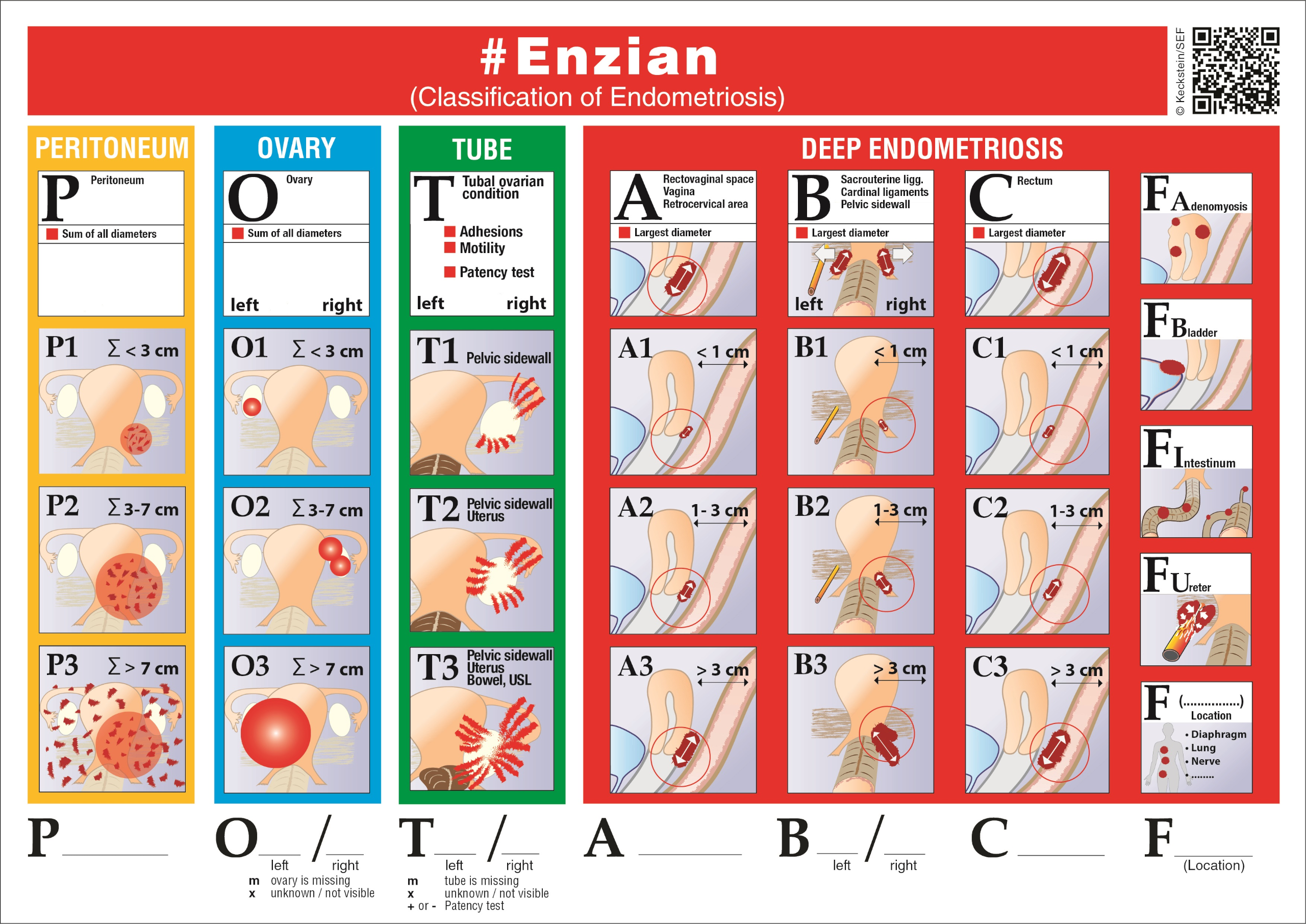
The different compartments (peritoneum, ovary, tube, TIE) are arranged next to each other. Other affected anatomical structures are also listed in the right-hand column. The severity, the size of the lesion, (1-3) is arranged from top to bottom.
The extragenital forms of the disease are given the letter F, e.g. FU (li/re) = ureteral obstruction with congestion, FB = bladder endometriosis, etc.. The findings are documented in the coding formula POT ABCF.
The compartments are classified as follows:
P=peritoneum(diameter of the circle in which the areas of all visible foci can be fictitiously summed =Σ) P1=<3cm, P2=3-7cm, P3=>7
O= Ovary (diameter of the sum of all endometriosis cysts; each side assessed separately; left/right) O1=<3cm, O2=3-7cm, O3=>7
T= adhesions and tubo-ovarian findings (adhesions to the pelvic wall, uterus and bowel, tubal patency= +/-; each side assessed separately; left/right).
A/B/C = deep endometriosis
A= rectovaginal septum and retrocervical area, vagina – B (left/right) = parametrium, sacrouterine ligament – C= rectosigmoid)
F: FA= adenomyosis, FI= further intestinal foci > 16 cm from ano, FU= ureter, congestion, F(…)= defined other localizations
The classification can be applied intraoperatively or preoperatively using ultrasound diagnostics or MRI, and is annotated with the lower case letters: [(s)= Surgical – (u)= Ultrasound – (m)= MRI)
for example: #Enzian (u) “Sonographic” classification: sonography can be used to visualize the ovaries (O), compartments A,B and C, the uterus (FA), the bladder (FB) and the ureters(FU). Adhesions between the ovary, the pelvic wall, the uterus and the bowel can be described using dynamic ultrasound examination with the “sliding effect”, as defined by the IDEA (International Deep Endometriosis Analysis) expert group.
Not all of these methods allow a complete recording of all findings. For example, peritoneal lesions cannot be assessed sonographically and intramural intestinal lesions are difficult to assess by diagnostic laparoscopy alone. In this way, the different methods of assessing findings complement each other. For structures that cannot be assessed (e.g. adhesions), the respective compartment is coded with (x). Only the affected compartments are coded so that the extent of the disease and the affected organs can be identified more easily and quickly when reading.